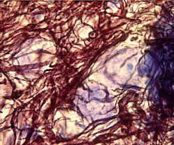
Neuron stains
I just got back from a neat lab for my hearing class, in which we got a tour of how to create 3-D models of neurons, see tonotopy in the cortex (with Fos labeling of cells stimulated at particular frequencies), and different types of stains of neurons in the cochlear nucleus.
Joe C. Adams showed us slides demonstrating the various neuron stains in a teaching microscope that had four extra eyepieces connected to it so that all five of us could look at the slides at the same time.
These pictures don’t do the staining justice. The Protargol (reduced silver) stains, which highlight nerve fibers, were so pretty that I wanted to make big prints of them and hang them on my wall. And I was hoping Google would help me find some really beautiful images of these things, but, alas.
I also got a kick out of seeing cartwheel cells, which look like cell bodies with arms coming out in all directions, and when you zoom in and out of the slide, you see new arms coming in and out of focus, making the whole cell look as though it’s spinning. Very cute!
One Comment
- jenny replied:
i have now set this:
http://researchpath.hitchcock.org/SocForHeme/casesQuarter/SaidAug96/sochem9634re50640.jpeg
as my desktop image.April 6th, 2007 at 1:49 pm. Permalink.
